What Is Skeletal Survey X-ray Examination: Bone Age?
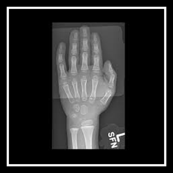
- Skeletal survey X-ray examination: Bone Age is a survey done to find out the bone age to assist the doctors to estimate the maturity of the skeletal system or the bones of children.
- This examination is usually done by taking the X-ray of the left wrist joint, hand and finger. It’s a fast examination, not painful and only involves low radiation dose. (http://kidshealth.org/parent/system/medical/xray_bone_age.html).
 Picture 1: Image of skeletal survey X-ray examination: Bone Age left hand
Source : http://kidshealth.org/parent/system/medical/xray_bone_age.html
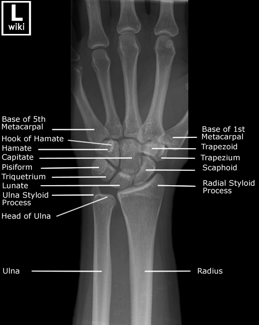
 Picture 2 : Anatomy of the left hand and wrist joint
Source : http://www.wikiradiography.net/page/Wrist+Radiographic+Anatomy
How And Where Can I Get This Examination?
- When you visit your doctor, he/she will decide if you require the examination.
- If required, the doctor will make a request for the examination using the Radiology Examination Request Form.
- This examination is available in MoH hospitals.
When Is Skeletal Survey X-ray Examination: Bone Age Required?
Skeletal survey helps in determining how fast or slow the growing or maturing process of a child’s bone compared to the increase in age. It assists doctors to diagnose factors affecting the fast or slow physical development of the child. This examination will be usually requested by pediatrician for the following reasons:
- Determine duration for a child to achieve growth
- Determine when the child will attain puberty.
- Determine the maximum height that can be attained by the child.
This survey is also usually used to monitor the development and as a reference for a child who has a growth defect that involves:
- Diseases that have hormonal effect on growth.
- Lack of growth hormone.
- Hypothyroidism.
- Premature puberty.
- Disruption of adrenal gland.
- Disruption of genetic growth such as Turner syndrome.
Before the examination
- The radiographer will explain to the patient about the examination. Accompanying person is allowed to listen to the radiographer’s explanation.
- Make sure you or the accompanying person are not pregnant or suspected to be pregnant. Please inform the radiographer if you are pregnant or suspected to be pregnant.
- Patient need to take out metal object such as watch and bangles on the hand to avoid image artifact.
- Radiographer will also explain briefly how the procedure is done.
During the examination
- Patient will be given a lead gown to wear for radiation protection.
- The left hand will be placed on top of a device to capture the X-ray image.
- Patient is advised not to move during the examination.
- Radiation protection will be provided.
After the examination
- No special after care is required.
- Patient will be allowed to leave after the examination.
- If you have any problem, please inform the radiographer.
Examination Report
All images produced will be reviewed by radiologist and report will be prepared.
References
- http://kidshealth.org/parent/system/medical/xray_bone_age.html
- http://www.wikiradiography.net/page/Wrist+Radiographic+Anatomy
| Last Review | : | 26 July 2017 |
| Writer | : | Mary Oommen |
| Accreditor | : | Daud bin Ismail |


