What Is CT Cardiac?
- CT Cardiac examination is performed to assess the risk of heart disease using the CT scan scanner.
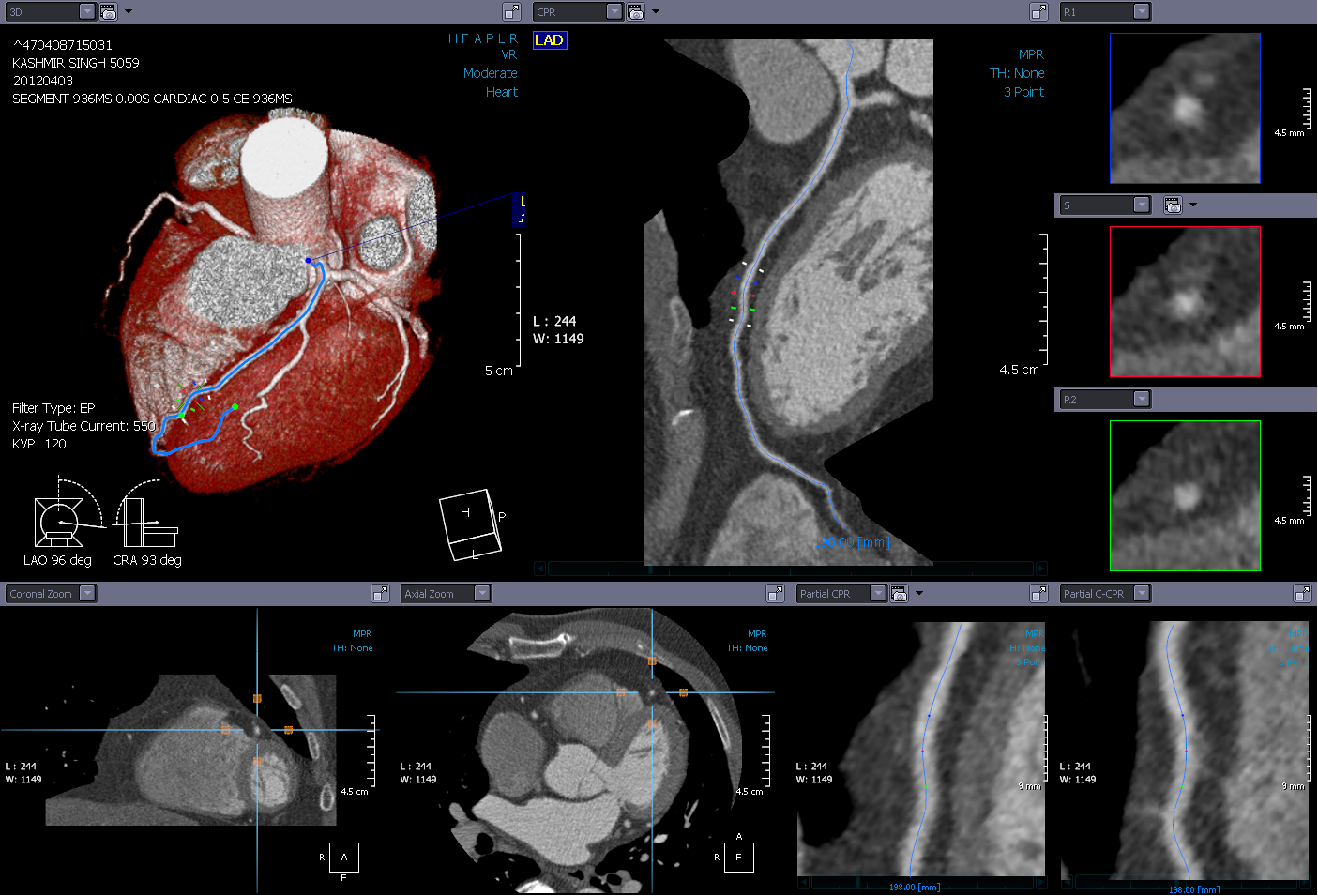
- A stable and low heart rate is important to allow the image of the heart to be visualised clearly as seen in the Figure 3.
- Contrast medium is used to visualise the blood vessels.
- CT Cardiac examination is a relatively safe and fast procedure.
Reasons For A CT Cardiac Examination
Some of the main reasons for CT Cardiac examination is to visualise and study the vessels of the heart. Normally CT Cardiac is performed because of: (Shakespeare C., 2014):
- You are experiencing pain/ tightness in your chest
- Hypertension
- Family history of heart disease
- Still experiencing chest pain after heart surgery
How And Where Can I Get This Examination?
This examination can only be requested by a specialist and performed at hospitals equipped with CT facilities.
Before examination
- Inform your doctor or radiographer if you are pregnant or suspect that you may be pregnant.
- Avoid drinks that contain caffein a day before the examination.
- You need to take steroids, if you have previous allergies or have had asthma.
- You will need to take medications to stabilise and reduce the heart beat (e.g : Metoprolol/ Beta Bloker).
- You have to fast 4 – 6 hours before examination.
- A needle (branula) will be inserted into the vein.
- You need to undress or accessories from your body and change to hospital gown.
- Your weight. Heart beat and blood ppressure will be taken prior to the examination.
- A stable heart rate is necessary to ensure successful examination.
During examination
- You will be instructed to lie on the examination couch.
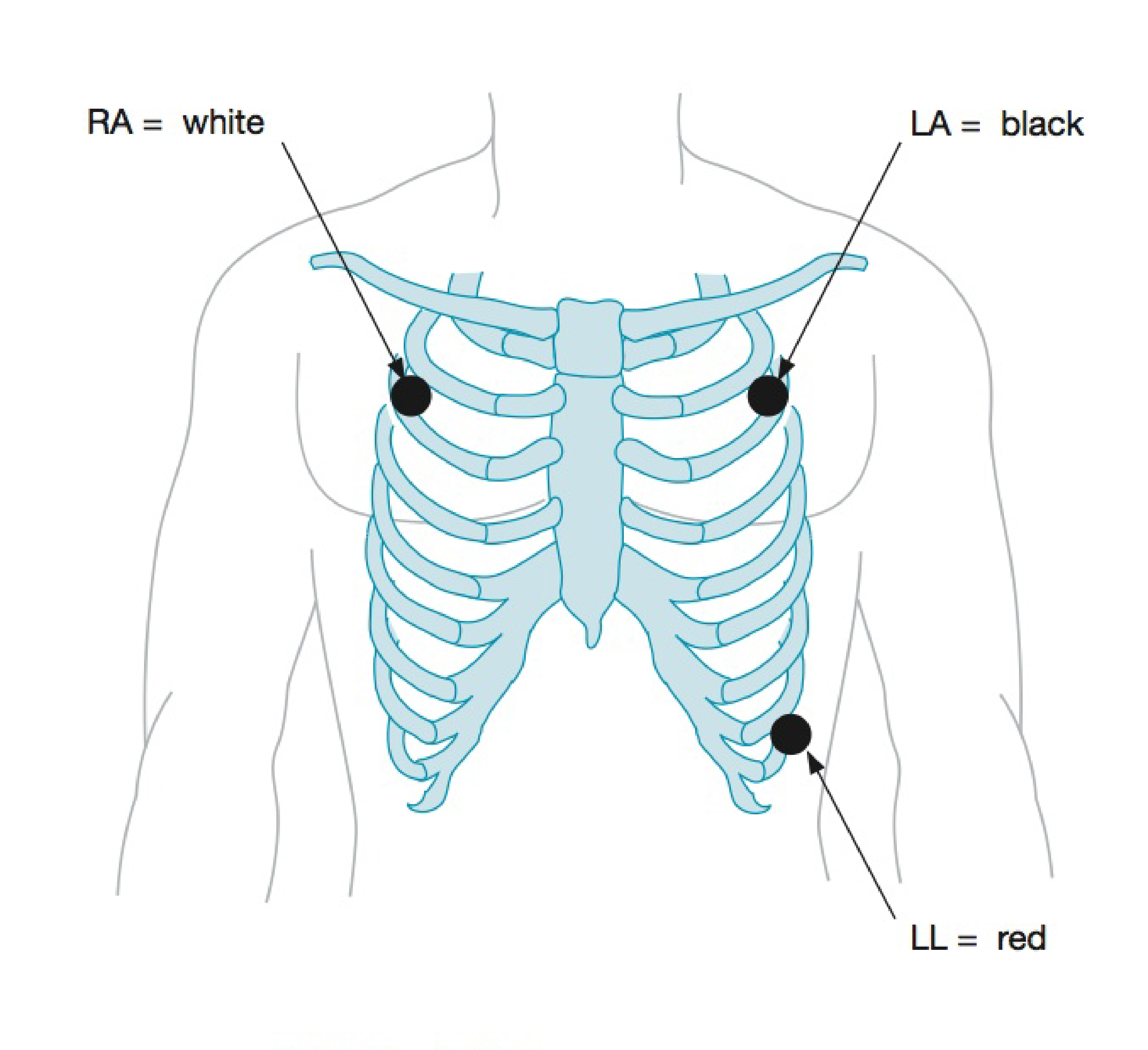
- The radiographer will attach the ECG (Electrocardiogram) leads on your chest ( Figure 1).
- The radiographer will give clear intsruction of the examination That you need to follow.
- During the scanning, the patient is allowed to communicate with the radiographer via intercom.
- The radiographer will place the GTN tablet (Glyceryl Trinitrate) under your tongue to expand the blood vessels.
- Contrast media will be injected into the vein by the doctor.
- Contrast media will be injected into the vein by the doctor.

Figure 1: ECG lead location placed on the patients’ chest
Source: http://lifeinthefastlane.com/education/procedures/lead-positioning/
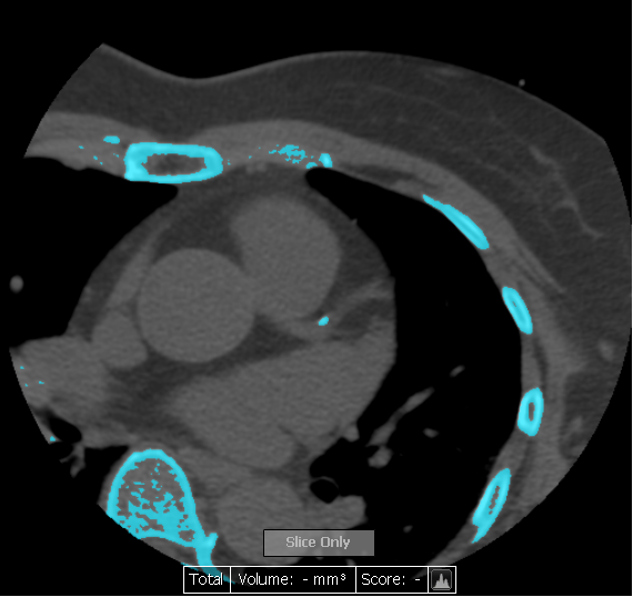
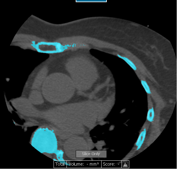
Figure 2 (a), (b): Calsium Score
Source: National Cancer Institute
Source: Hospital Raja Permaisuri Bainun
After examination
- The heart beat and blood presure will be monitored before you are allowed to go home.
- On completion of the examination, you are adviced to follow the appointment given by the requesting clinic.
- You are allowed to eatand drink plenty of water to speed up the contrast media excretion of the body through the urine.
Examination report
All images that are produced are reviewed by the radiologist and report is prepared.
Reference
- Cadogan M, 2014, Lead positioning, 3-electrode system, Life In The Fast Lane, (http://lifeinthefastlane.com/education/procedures/lead-positioning/)
- Shakespeare C, Kardiak CT Angiography, 2014, (http://www.kardiakctangiography.com/ use_kardiak_ct_angiography.htm)
| Last Review | : | 24 July 2017 |
| Writer | : | Pushpa Thevi Rajendran |
| Accreditor | : | Daud bin Ismail |


