What is X-Rays?
X-rays is a form of radiation, just like visible light or radio waves. Unlike visible light, X-rays can penetrate human body. When these devices are used appropriately with proper techniques, X-rays is able to produce an images of the internal structures within human body. These images can be viewed on a photographic film or on a TV or computer screen. X-ray examinations is useful in providing valuable information on human health and play an important role in assisting your doctor to make a more accurate diagnosis. In some cases X-rays is used as a guiding tool when placing tubes or other devices in the body or with other therapeutic procedure.
Production of X-Ray Image
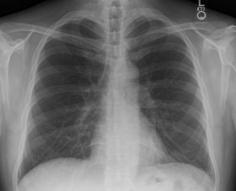
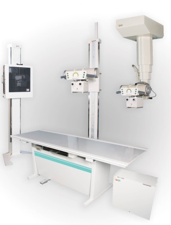
A special machine is used to emit a small amount of ionising radiation known as X-rays. This radiation will then passes through your body and falls on a film or similar device to produce the image. Nowadays X-ray examinations uses a device that captures transmitted X-rays to create an electronic image. The calcium from bones will prevent the passage of radiation. Therefore, in healthy person the bones are seen as white or grey objects. On the other hand, radiation passes easily through an air spaces. That is why a healthy lungs appears as black objects.
Imaging Methods Using X-Rays
There are various imaging methods using an X-rays such as:
-
Fluoroscopy – also known as ‘screening’. Often used to monitor movement of internal organs, such as stomach, intestines, etc. It is also used to guide in biopsies or assisting fluid removal from chest and abdomen, aids in detecting presence of clogs or cholesterol in the arteries and arterioles of the blood vessels of the heart and brain. A life moving image can be seen on a TV screen. The radiologist and radiographer performing the examination can take a snapshots of any important findings or do recording of the whole scenario on a video. Fluoroscopic examinations usually uses higher radiation doses than simple radiography.
-
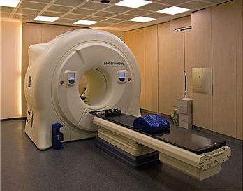
Computed Tomography (CT) Scan- this is a more sophisticated way of using X-rays. The computer will slice the images to create a three-dimensional (3D) pictures and displayed on a computer monitor. If many slices are imaged, the radiation dose can be as high as or higher than fluoroscopic examinations.
Performing X-Ray Examinations
X-ray examinations normally performed by a qualified radiographer and radiologist. In the dental practice the dental surgery assistant and registered dental practitioner is allowed to perform certain dental X-ray examination.
Benefits of X-Ray Examinations
The benefits it offers is to help doctors in making correct diagnosis, thus formulate the right regime of treatment, and this will outweigh any small risk involved. If treatment plan is solely based on clinical findings, then an exposure to radiation even though with small risk, might give a big impact to your health may likely to be greater.
Risk for Radiation Induced Cancer
The risk of induced cancer from radiation is known to be low but is additive. In every examination done, the risk to the patient is high. Thus, keeping doses to a minimum, adequate just for a diagnostic purposes with god quality images is therefore recommended. Perhaps, with this, the probability of one to developing cancer within 10 years from X-ray examination can be ignored (very small).
X-Ray Safety
X-ray examination is painless and is a non-invasive procedure. It does not cause any side effects. X-rays are safe when used with care. Radiologists and radiographers are trained and are able to manipulate minimum amount of radiation necessary with optimum result obtained. Proper handling of conducted imaging carries minimal risks and should be performed whenever is required from clinic. The amount of radiation used in most examinations is very minimal and yet, the benefits are greatly outweigh the risk of harm. X-rays are produced only when a switch is momentarily turned on. As with visible light, no radiation remains after the switch is turned off and you will not be radioactive after the test.
X-Ray Examinations Doses
Radiation dose is often measured using the quantity of effective dose, expressed in milisievert (mSv). The effective dose is a dose that is given to the whole body enough to cause risk of cancer when imparted to various organs in a specific part of the body. Effective dose offers a way to compare approximately the relative risk between different radiation procedures. Every person is at risk of being exposed to various radiation sources such as from surroundings, cosmic radiation, radiation from earth, food, and even our own body. This radiation (gamma rays) is similar to X-rays used in medical examinations. Geographically, regardless from where they come from, every individuals have the potential risk of being exposed to radiation in the range of 1 to 3 mSv every year. Each X-ray examination, imparts different amounts of radiation. The most common X-ray examination is the chest X-ray (frontal view). It imparts an average dose about 0.02 mSv.
Below are the recommended doses for common radiological examinations in an adult patient with average size.
| X-ray Examination | *Mean Effective Dose (mSv) | Number Equivalent Chest X-ray |
| Skull X-ray | 0.1 | 5 |
| Thoracic Spine/Lumbar Spine X-ray | 1.0 – 1.5 | 50 – 75 |
| Mammography | 0.4 | 20 |
| Pelvis/Hip/Abdomen X-ray | 0.6 – 0.7 | 30 – 35 |
| Knee/Other Extremities | 0.001 – 0.005 | 0.05 – 0.25 |
| Spine CT | 6 | 300 |
| Abdomen/Pelvis CT | 6 – 8 | 300- 400 |
| Head/Neck CT | 2 – 3 | 100 – 150 |
| CT Coronary Angiography | 16 | 800 |
| CT Virtual Colonoscopy | 10 | 500 |
The Effect of Having Many X-Rays
Various types of X-ray examination carries different amount of radiation dose. To estimate the effect of having many exposure from examinations, the dose for each X-ray examinations are simply added together. In the case that if you need a new X- ray examination and you are concern about the effect since you have been exposed to a large number of X-rays previously, then the need should be judged on its own merits. Before one could proceed, your doctor must be satisfied that there is no other way in gaining new information that is essential in providing effective management of your medical problem. Make sure your doctor is also aware of other X-rays or scans that you have had earlier to avoid unnecessary additional examinations.
Pregnant Women for X-Ray Examinations
The radiographer or radiologist should ask their female patients especially at the childbearing age if they are pregnant. If they are confirmed to be pregnant, then they need to further discuss the matter with their doctors either to proceed with the examinations or to recommend postpone. As long as the X ray examination helps and benefits clinical findings after all other available examination options had been discussed, the risk of radiation exposure which is very small shall just be ignored. Modern equipment with the help of good technique available today, examination of the head, feet, neck, shoulder and even chest can be safely carried out during pregnancy. As for other type of examination, special considerations may be required. Female patients should inform their physician and the healthcare provider concerned, about their pregnancy or even the possibility of being pregnant. Having been made aware of this condition, the physician or healthcare provider, in cooperation with the radiation protection specialist will then weight the benefits and risk in performing the abdominal and pelvic investigations, especially using relatively high dose procedures (CT and fluoroscopy).
Younger Patients for X-Ray Examination
There is no restrictions regarding use of X-rays to children, provided that clinical benefit exceeds the potential risks of radiation which is relatively small. Children may have organs that is more sensitive to higher radiation than adults and their radiation risks may be as twice as the risk among the middle-aged group of the same X-ray examination. This is because children have a longer life expectancy, so there is more time for a radiation-induced cancer to develop. This is why special attention is needed to ensure that every child who is X-rayed will be benefited. The radiation dose is also kept as low as possible without detracting any useful information the examination can provide. The use of other alternative imaging techniques that do not use ionizing radiation should always be considered. Radiological procedures to children should be individually planned and limited to what is thought sufficient for a correct diagnosis.
Radiation Risks for Future Generations
If the reproductive organs (ovaries and testes) are exposed to radiation there is a possibility that heredity diseases or abnormalities may passed on to future generations. Although such effects has never shown in humans, yet a lead-rubber shields is used and placed over the reproductive area during X-ray examinations, as a precaution step. These lead-rubber shields are only necessary for examinations in the lower abdomen and thighs area especially on young patients. However, in some instances, these lead-rubber shields such as gonad shields is not needed since they will obscure important diagnostic information.
Alternatives to X-Ray Examinations
Although most studies have shown that the risk with single X-ray is very small, it is a question of how to minimise the risk. Depending on medical requirement, alternatives to X-ray examinations may be necessary and these include:
- Ultrasound- the use of sound waves to create an image of internal body structure.
- Magnetic Resonance Imaging (MRI)- the combination of magnetic field and radio waves to produce 3D pictures
Unlike X-rays, these procedures are not known to increase the risk for cancer. However it is not always possible to replace X-ray with a non-ionizing radiation examinations since there are many other things that need to be considered such as the need for sedation in young children for MRI as opposed to a CT examination.
References
- X-ray Examinations, www.betterhealth.vic.gov.au, Copyright ©1999/2014 State of Victoria
- National Radiological Protection Board, X-rays How Safe Are They?, May 2001, UK
- Kementerian Kesihatan Malaysia, Pekeliling Keperluan Mendapatkan Khidmat Juru X-ray Berkelayakan Sebagai Pengendali Radas Penyinaran Bagi Klinik Pengamal Perubatan dan Institusi Perubatan Swasta Di Bawah Akta Perlesenan Tenaga Atom 1984 (Akta 304) Bagi Maksud Perubatan, 2010, Putrajaya
- Kementerian Kesihatan Malaysia, Pekeliling Mendapatkan Khidmat Personel Berkelayakkan sebagai Pengendali Radas Penyinaran Bagi Perkhidmatan Pergigian Di Bawah Akta 304 Bagi Maksud Perubatan, 2011, Putrajaya
- Patient Safety: Radiation Dose in X-ray and CT Exams, wwww.radiologyinfo.org, Copyright © 2014 Radiological Society of North America, Inc
- RPOP Website: http://rpop.iaea.org and FA Mettler et al, Radiology 008; 248:254-63
| Last Reviewed | : | : 8 October 2014 |
| Writer / Translator | : | Suratey bt. Sulaiman |
| Accreditor | : | Nik Mohamed Hazmi b. Nik Hussain |


