What Is Knee X-ray Examination?
- Examination done by a radiographer on the knee joint.
- It’s not painful.
- The knee X-ray examination includes parts of knee bone, together with the thigh bone (the bone above knee), tibia and fibula (the bones below knee), patella (knee cap) and soft tissues.
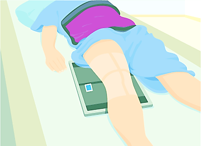
Picture 1: Illustration shows one of the positioning technique for knee X-ray.
Source: http://kidshealth.org/parent/system/medical/xray_knee.html#
How And Where Can I Get This Examination?
- When you visit your doctor, he/she will decide if you require the examination.
- If required, the doctor will make a request for the examination using the Radiology Examination Request Form.
- This examination is available in MoH hospitals and health clinics with diagnostic imaging service.
When Is Knee X-ray Required?
- Knee X-ray examination helps to find the causes and signs of pain, swelling, sprain, or abnormalities of the knee. It can also detect fracture of bones and dislocation of the joints.
- In a knee surgery, knee X-ray will be taken before the surgery for planning the surgery and also after surgery for checking.
Before the knee X-ray examination
- You are advised not to bring or wear any valuables.
- Children can be accompanied by an adult family member.
- Make sure you are not pregnant or suspected to be pregnant. Please inform the radiographer if you are pregnant or suspected to be pregnant.
- You will be briefed about the procedure.
- You may need to change into hospital attire.
During the examination
- Two basic projections usually done are:
– Anteroposterior (AP) – to visualize the knee joint and the front part.
– Lateral (X-ray done from the side of the knee joint).
- Usually the examination is done by lying down or standing.
- Radiation protection device will be provided during examination.
- This examination takes approximately 10 minutes only.
- Additional projections will be done if required.
After examination
You will be allowed to leave after the examination.
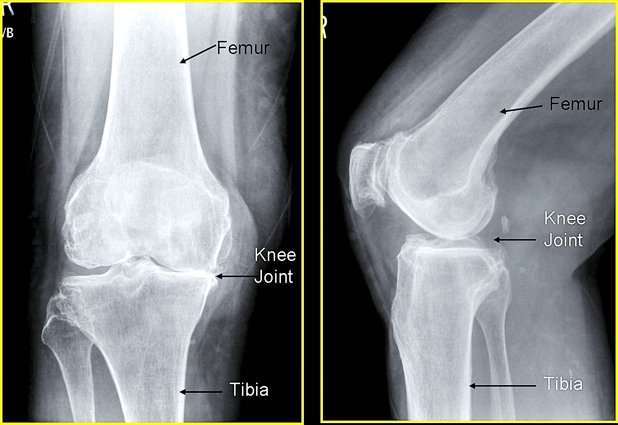
Picture 2: Example of a normal knee X-ray image.
Source: http://orthoanswer.org/foot-ankle/fracture-foot-ankle/investigations.html
Examination Report
- All images produced will be reviewed by radiologist and report will be prepared.
- Examination result will be sent to the doctor seeing you.
References
- http://kidshealth.org/parent/system/medical/xray_knee.html#
- http://www.footvitals.com/injuries/foot-xray.html
- http://radiology-anatomy.blogspot.com/2013/04/knee-joint-x-ray-anatomy.html
- http://orthoanswer.org/foot-ankle/fracture-foot-ankle/investigations.html
| Last Review | : | 31 July 2017 |
| Writer | : | Mary Oommen |
| Accreditor | : | Daud bin Ismail |