Introduction
Cyst is defined as an abnormal cavity in hard or soft tissue which contains fluid, semifluid or gaseous. Cyst is often encapsulated and lined by epithelium.
An oral cyst is generally filled with liquid. Cyst can form anywhere in the mouth, including the lips, tongue, floor of mouth, gums, palate, throat, salivary glands or cyst can occur in the soft tissue and may appear as a small, painless bump. It is often less than 1 cm in diameter. If infected it becomes red, swollen and painful. It can also occur in the bone where a large oral cyst inside the bone can weaken the jaw. Most cysts of the jaws arise from odontogenic epithelium (which forms the teeth). The lower jaw is affected three times as commonly as the upper jaw.
The Types of Cysts
Cysts are classifed into inflammatory cysts or developmental cysts.
Inflammatory cysts are the most common of all jaw cysts and arise is associated with non-vital tooth include the following:
- Radicular Cyst (Periapical) – It develops as a result of an infection in the tooth pulp. Most common cyst found in the mouth.
- Residual Cyst – Periapical cysts remains after the removal of the non-vital tooth from which it arose.
Odontogenic keratocyst which was previously classified as inflammatory cyst is now considered an odontogenic tumour.
Developmental cysts include the following:
- Dentigerous cyst –
Associated with unerupted tooth. They develop in the dental follicle and are often unnoticed because they cause no pain. It can displace the unerupted tooth or adjacent teeth. Usually involves the lower wisdom tooth and upper canine tooth.
- Eruption Cystsconsidered as minor soft tissue forms of dentigerous cysts. Often burst spontaneously.
- Soft tissue cyst
Mucocele – occurs when the salivary duct ruptures,usually due to trauma. Painless growth, tend to rupture on their own and often heal quickly without any intervention. Continue to increase in size if it does not rupture. Organize and form a bump on the inner surface of the lip. It is called ranula when occurs on the floor of the mouth
Clinical Presentation
Signs of cyst
Oral cyst is often asymptomatic and is usually an incidental finding on radiograph which shows radiolucency. It is slow growing and may reach a large size before giving rise to symptoms such as:
- Bone expansion/swelling (slow-growing) with normal overlying mucosa.
- Discharge: usually into the mouth
- Pain: due to secondary infection
- Ill fitting denture
- Displaced teeth or missing tooth in normal series
- Distortion of adjacent structures
- Mobile teeth/ Non vital tooth
Implication
Secondary effects on jaw due to oral cyst are jaw asymmetry, pathological fracture of jaw, secondary infection and numbness (if compressing on nerve).
Diagnosis is based on history, clinical examination and investigation such as pulp vitality testing, radiograph (intraoral and extraoral), aspiration and analysis of cyst fluid and histopathological examination (biopsy).
Management
Management of cyst is removal by surgery by enucleation or marsupiallization or surgical excision. Enucleation is thecomplete removal of the cyst lining is usually done under local anesthesia for small lesions and under general anaesthesia for bigger size oral cyst.
Marsupialization is the partial removal of the cyst. This is a type of decompression procedure for the treatment of bigger cysts to reduce the size of the cyst. A window or fenestration is made in the bone and the cystic contents are evacuated. The cyst lining is left behind. The hollow cavity is then packed till it gets obliterated by bone slowly over a period of time. Antibiotics may be given if the cyst is infected.
Removal Of Cyst By Surgery
|
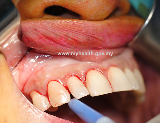
1. Cyst at area of anterior maxilla. Incision line follow gingiva margin
|
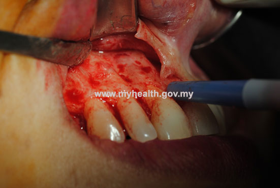
2. Flap was raised to expose cyst.
|
|
|
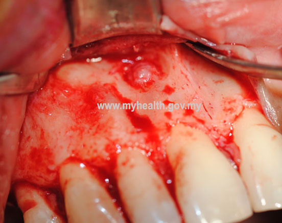
3. Cyst were exposed and enucleated
|
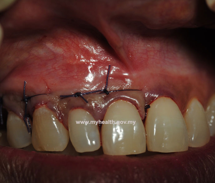
4. Flap reposition and sutured.
|
|
|
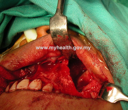
5. Big cavity after cyst removal
|
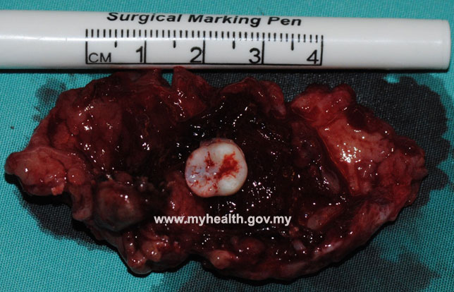
6. Cyst lining and tooth attached to it (Dentigerous cyst)
|
|
|
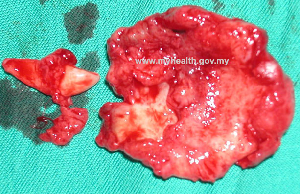
7. Cyst lining and tooth related to cyst (Dentigerous cyst)
|
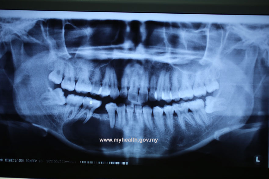
8. X-ray shows well margin of cyst.
|
References
- Oral and Maxillofacial Medicine. The Basis of Diagnosis and Treatment. Second Edition. Churchill Livingstone. Crispian Scully
- Oral and Maxillofacial PATHOLOGY. Second Edition. Elsevier. Neville Damm, Allen Bouquot
| Last Reviewed | : | 7 August 2014 |
| Writer | : | Dr. Mohd Rosli b. Yahya |
| Accreditor | : | Dr. Kok Tuck Choon |


