Introduction
The oral mucosa can take up a variety of discolorations, including brown, blue, gray and black. Such color changes are often attributed to the deposition, production, or increased accumulation of various exogenous (extrinsic) and endogenous (intrinsic) pigmented substances.
Extrinsic Mucosal Discoloration
Extrinsic discolouration is superficial and usually caused by:
- Habits like tobacco or betel use (Figure 1)
- Coloured foods or drinks (coffee, tea, beetroot, liquorice)
- Drugs (chlorhexhidine, iron salts, minocycline, lansoprazole)

Figure 1: Brown discoloration of oral mucosa and generalized black discoloration of teeth due to chronic betel quid chewing. (Oral Medicine Clinic, HRPB, Ipoh) - Black hairy tongue
- One of extrinsic type of discolouration seen especially in patients on a soft diet, smokers and those with dry mouth or poor oral hygiene.
- To overcome this problem, it is best to avoid the cause where known, and to advise the patient to brush the tongue or use a tongue-scraper.

Figure 2: Black hairy tongue (From Scully & Felix, 2005)
Intrinsic Staining
Hyperpigmentation is caused by increased in melanin or number of melanocytes, or other materials.
It can be divided into:
- Localised
- Multiple or Generalised
Localised Mucosal Intrinsic Hyperpigmentation
Causes included:
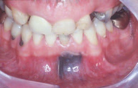
- Amalgam tattoo (embedded amalgam) – a single blue-black macule usually in the mandibular gingiva close to the scar of an apicectomy or where amalgam has accidentally been introduced into a wound. It is painless, and does not change in size or colour.
- Graphite tattoo – may be seen where a pencil lead has broken off in mucosa.
- Naevi– blue-black often papular lesions formed from increased melanin – containing cells (naevus cells) seen particularly on the palate.
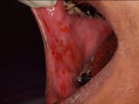
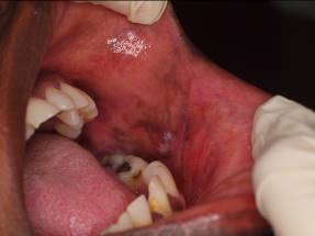
- Pigmentary incontinence (local irritation/inflammation) – may be seen in some inflammatory lesions such as lichen planus, especially in persons with pigmented skin (dark skin).
- Melanotic macules – are usually flat single brown, collections of melanin-containing cells, seen particularly on the vermilion border of the lip and on the palate. Mostly smaller than 1 cm, painless and do not change rapidly in size. Seen mainly in white people.
- Neoplasm. E.g:
- Malignant melanoma– rare, seen usually in the palate or maxillary gingivae. About one-third arise in areas of hyperpigmentation. Features suggestive of malignancy include a rapid increase in size, change in colour, ulceration, pain, the occurrence of satellite pigmented spots or regional lymph node enlargement.
- Kaposi’s sarcoma– usually a purple lesion seen mainly in the palate or gingival of HIV-infected and other immunocompromised persons.
- Pigmented neuroectodermal tumour of infancy– a rare, rapidly growing, pigmented benign neoplasm of neural crest origin, primarily affects the maxilla of infants during the first year of life.

Figure 3: Amalgam tattoo (From Scully & Felix, 2005)
Figure 4: Pigmentary incontinence/post-inflammatory hyperpigmentation in lichen planus.
(Oral Medicine Clinic, HRPB, Ipoh)
Multiple or Generalised Mucosal Intrinsic Hyperpigmentation
Causes included:
- Genetic. E.g:
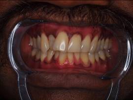
- Racial pigmentation – Usually in dark-skin people, mainly on the gingivae.
- Carney syndrome- Autosomal dominant syndrome with presence of myxomas, spotty lip pigmentation and endocrine overactivity.
- Laugier-Hunziker syndrome- Labial and oral mucosal brown to black pigmented macules and nail hyperpigmentation.
- Lentiginosis profusa and Leopard Syndrome- allelic disorders caused by mutation in PTPN11, a gene encoding the protein tyrosine phosphatase SHP-2, with many abnormalities including multiple lentigines (flat tan, brown or black spots, also called liver spots)
- Peutz-Jeghers syndrome- an autosomal-dominant condition due to a chromosome 19 gene mutation with circumoral melanosis and intestinal polyposis. Oral brown or black macules appear in infancy and affect especially the lips, buccal mucosa and may be seen on extremities and abdomen.
- Café au lait pigmentation- tan or brown coloured, irregularly shaped macules of variable size. May occur on the skin and unusual examples of similar appearing oral macular pigmentation have been described in some patients. May be identified in a number of different genetic diseases, including neurofibromatosis type I, McCune-Albright syndrome and Noonan’s syndrome.

Figure 5: Racial pigmentation (Oral Medicine Clinic, HRPB, Ipoh)
- Drugs. E.g:
- ACTH, zidovudine- may produce brown pigmentation
- Antimalarials- produce a variety of colors in mucosa, ranging from yellow with mepacrine to blue-black with amodiaquine.
- Busulphan, some other cytotoxic drugs, contraceptive pill, phenothiazines and anticonvulsants- may occasionally produce or increase brown pigmentation
- Minocycline- may cause blackish discoloration of the teeth, gingivae and bone, skin, sclera and even breast milk.
- Tobacco- smoker’s melanosis
- Endocrin. E.g:
- Addison’s disease (hypoadrenalism)
- May cause generalized or patchy hyperpigmentation due to excessive production of ACTH, which has similar activities to MSH (melanocyte stimulating hormone).
- Usually autoimmune (idiopathic), but hypoadrenalin may be seen in HIV disease.
- Pigmentation usually generalized, but most obvious in normally pigmented areas (eg the nipples, genitalia), skin flexures, and sites of trauma.
- The oral mucosa may show patchy hyperpigmentation.
- Patients also typically have weakness, weight loss, and hypotensio
- Addison’s disease (hypoadrenalism)
-
- Nelson syndrome – a rare condition caused by ACTH-overproduction in response to adrenalectomy, usually for Cushing disease or breast cancer. It is a spectrum of symptoms and signs arising from an ACTH – secreting pituitary macroadenoma after a therapeutic bilateral adrenalectomy. Hyperpigmentation of the skin is usually obvious and is not limited to sun-exposed areas. The degree of pigmentation varies depending on the racial origin of the patient and the serum concentrations of ACTH. Patients usually appear hyperpigmented with a linea nigra (pigmentation extending up the midline from the pubis to the umbilicus) and pigmentation of scars, gingivae, scrotum and areolae. Other signs and symptoms related to the pituitary tumour itself like hypopituitarism, headache and visual disturbance are also a common presentation
-
- Pregnancy – Pathogenesis is not really understood, however it is considered to rely on increased serum levels of melanocytic-stimulating hormone, estrogens, and possibly progesterone, which stimulate melanocytic activity contributing to pigmentation. Facial pigmentation including melasma, chloasma or mask of pregnancy may affect up to 70% of pregnant women. The most common is centrofacial melasma developing on the forehead, cheeks, upper lip, and chin. Both pregnancy and use of oral contraceptive have also been associated with oral mucosal melanosis.
-
- ACTH-producing tumour (e.g. bronchogenic carcinoma) – may induce brown pigmentation, particularly of the soft palate.
Diagnosis
Patients with generalized or multiple hyperpigmentation:
- To exclude Addison’s disease:
- Blood pressure (hypotension)
- Plasma cortisol levels (low level)
- Synacthen test (ACTH test) (impaired response to adrenocortitrophic hormone)
- Specialist referral is indicated.
Patients with Localized hyperpigmentation:
- Radiographs – can show amalgam, graphite or foreign body, or bone rarefaction in pigmented neuroectodermal tumour of infancy.
- Photographs – for future comparison of size and colour.
- Urinary cathecolamines- raised in pigmented neuroectodermal tumour
- Biopsy (to exclude melanoma) – particularly where there is a solitary raised lesion, a rapid increase in size, change in colour, ulceration, pain, evidence of satellite pigmented spots or regional lymph node enlargement.
Management
Management is of the underlying condition.
References
- Scully C 2008. Oral and Maxillofacial Medicine: The Basis of Diagnosis and Treatment. Elsevier Health Sciences. Page 114-8
- Scully C, Felix DH 2005. Oral medicine- update for the dental practitioner: 6. Red and pigmented lesions. Br Dent J 199:639-45
- Aguirre A, Alawi F, Luis Tapia J 2015. Pigmented lesions of the oral mucosa in Glick M, Burket’s Oral Medicine, 12th edition. People’s Medical Publishing House-USA. Page 123-46.
- N.N. Andrade et al. 2016. Melanotic neuroectodermal tumour of infancy – A rare entity. Journal of Oral Biology and Craniofacial Research 6 (2016) 237-40
- Dermatological Manifestations Associated With Pregnancy: Pigmentary Changes. http://www.medscape.org/viewarticle/706769_3
| Last Reviewed | : | 14 June 2017 |
| Writer | : | Dr. Khamisah bt. Awang Kechik |
| Accreditor | : | Dr. Ajura bt. Abdul Jalil |


