Introduction
A pleural biopsy is a procedure in which a sample of the pleura (the membrane that surrounds the lungs) is removed with a special biopsy needle to determine if infection, cancer, or another condition is present.
Conventional or closed pleural biopsy
Indications for pleural biopsy are to:
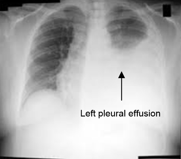
- Evaluate an abnormality of the pleura seen on chest X-ray (Figure 1)
 |
| Figure 1. Chest radiograph showing left pleural effusion |
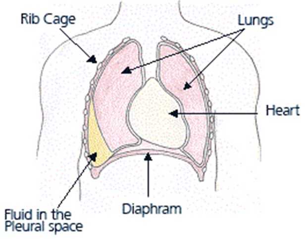
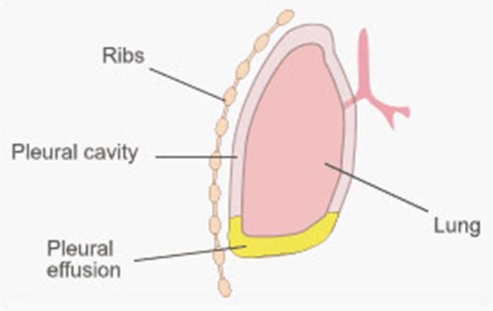
- Investigate pleural effusion (fluid collection in the pleural space) (Figure 2a & 2b)
- Diagnose the cause of a pleural infection (caused by bacteria, virus or fungus) or other condition
- Determine if a pleural mass is malignant (cancerous) or benign
- Obtain further information after pleural fluid analysis suggests the presence of cancer, tuberculosis or other infection
 |
| Figure 2a : Right pleural effusion |
 |
| Figure 2b |
Contraindications:
The absolute contraindication is any uncorrectable bleeding condition
Relative contraindications are:
- Inability to drain fluid from the pleural space (dry tap)
- Presence of pus in the pleural cavity
- An uncooperative patient
- High blood urea level
Patient preparation pre-procedure:
- Explanation about the procedure is given by the doctor to the patient.
- The doctor will ask if the patient has allergy to any medication, latex or tape
- A consent is required
- Generally, fasting is not required before the procedure
- If the patient is taking medications such as aspirin, blood thinning medications or other medications that affect blood clotting, it may be necessary to stop or replace these with other types of medications
- An ultrasound to localize the site of pleural biopsy is done
During the procedure:
The patient will be asked to:
- Remove any jewelry or other objects that may interfere with the procedure.
- Remove clothing, and will be given a gown to wear.
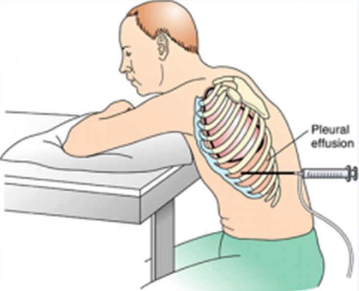
- Sit with arms raised and resting on a table (Figure 3). This position aids in spreading out the spaces between the ribs for needle insertion. If the patient is unable to sit, he/she may be placed in a side-lying position on the edge of the bed on the unaffected side.
- Hold still, exhale deeply, and hold his/her breath when told to during the procedure.
 |
| Figure 3. Position during pleural biopsy |
The following are done/given:
- Supplemental oxygen as needed, through a face mask or nasal cannula (tube).
- The skin at the puncture site will be cleansed with an antiseptic solution.
- A local anesthetic will be administered at the site where the biopsy is to be performed.
- A needle will be inserted between above the ribs.
- Once the pleural space is entered with the needle, fluid may be withdrawn slowly.
- A biopsy needle (Figure 4) will inserted into the site. One or more samples of pleural tissue will be obtained.
- The biopsy needle will be withdrawn and firm pressure will be applied to the biopsy site for a few minutes until any bleeding has stopped.
- A sterile adhesive bandage or dressing will be applied.
- The tissue sample will be sent to the lab for examination.

 |
| Figure 4. Abrams pleural biopsy needle (A – needle, B – stylet) |
Post-procedure:
- Dressing over the puncture site will be done
- Dressing will be monitored for bleeding or other drainage.
- A chest X-ray taken is after the biopsy.
- The patient may resume usual diet and activities if there are no complications
Monitoring of:
- Vital signs (heart rate, blood pressure, breathing rate, and oxygen level) are done before, during and after the procedure
Complications
The following may be the complications of a pleural biopsy
- Pneumothorax. Collapse of the lung, causing air to become trapped in the pleural space.
- Bleeding in the lung
- Infection
References
- Salyer WR, Eggleston JC, Erozan YS. Efficacy of pleural needle biopsy and pleural fluid cytopathology in the diagnosis of malignant neoplasm involving the pleura. Chest. 1975 May. 67(5):536-9. [Medline].
- Frank W. [Current diagnostic approach to pleural effusion]. Pneumologie. 2004 Nov. 58(11):777-90. [Medline].
- Prakash UB, Reiman HM. Comparison of needle biopsy with cytologic analysis for the evaluation of pleural effusion: analysis of 414 cases. Mayo Clin Proc. 1985 Mar. 60(3):158-64. [Medline].
- Koss MN, Fleming M, Przygodzki RM, Sherrod A, Travis W, Hochholzer L. Adenocarcinoma simulating mesothelioma: a clinicopathologic and immunohistochemical study of 29 cases. Ann Diagn Pathol. 1998 Apr. 2(2):93-102. [Medline].
- Christopher DJ, Peter JV, Cherian AM. Blind pleural biopsy using a Tru-cut needle in moderate to large pleural effusion–an experience. Singapore Med J. 1998 May. 39(5):196-9. [Medline].
- James P, Gupta R, Christopher DJ, Balamugesh T. Evaluation of the diagnostic yield and safety of closed pleural biopsy in the diagnosis of pleural effusion. Indian J Tuberc. 2010 Jan. 57(1):19-24. [Medline].
- Gouda AM, Al-Shareef NS. A comparison between Cope and Abrams needle in the diagnosis of the pleural effusion. Annals of Thoracic Medicine. 2006. 1(1):12-5.
- Scerbo J, Keltz H, Stone DJ. A prospective study of closed pleural biopsies. JAMA. 1971 Oct 18. 218(3):377-80. [Medline].
- Pandit S, Chaudhuri AD, Datta SB, Dey A, Bhanja P. Role of pleural biopsy in etiological diagnosis of pleural effusion. Lung India. 2010 Oct. 27(4):202-4. [Medline]. [Full Text].
| Last Reviewed | : | 13 November 2015 |
| Writer | : | Dr. Norhaya bt. Mohd Razali |
| Accreditor | : | Dr. Jamalul Azizi b. Abdul Rahaman |


